Being able to see how the brain grows, changes and sometimes malfunctions at the cellular level could help scientists improve their understanding of the many neurological disorders that reduce the quality of life for one third of the world’s people.
“Like a detailed GPS for the complex landscape of the brain, the brain atlas serves as an essential reference tool,” said Katrin Amants, a neuroscientist at the Jülich Research Center in Germany, who supervised Europe’s leading human brain mapping project, EBRANS.
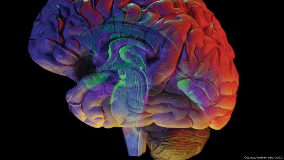
published in journal Nature, A new set of brain atlases There is hope to build on the EBRAINS project. It details how the brain develops in humans and other mammals from an early stage of cell division into a rich and diverse assortment of specialized cells that perform specialized functions.
Amants was not involved in the new research, but he told DW that the latest brain atlas, which includes others in development around the world, could lead to better diagnostic techniques for disorders such as Alzheimer’s or epilepsy. Some atlases can be used to study neurodevelopmental conditions such as autism or autoimmune complications such as multiple sclerosis.
Other benefits may include “more precise neurosurgery planning, and targeted treatments such as deep brain stimulation for Parkinson’s disease,” Amants said in an email.
“By providing a spatial blueprint across species, they help translate research into treatments that may one day improve the quality of life for millions of people.”
Several maps give a glimpse of our gray matter
What’s unique about the new brain atlases is that they show more than a single-point snapshot of cells in the brain, but how they change over time.
Officially, they only draft brain maps, but “the insight they provide is phenomenal,” Amants said.
The atlas is composed of data from human, non-human primate, and mouse studies. In total, 12 studies from research centers in North America, Sweden, Belgium and Singapore form the core of the work.
“The idea is to take all these snapshots at different times and you try to stitch them together to create this map of changes,” said Hongkui Zeng, director of the Allen Institute for Brain Science in the US.
Zeng’s research group monitored early gene signatures in developing cells and tracked how these structures change and move over time in the mouse brain to create a “trajectory map.”
“Once we have this map, we can take a diseased tissue, and we do the same thing, we create the profile and we match the cells to the reference atlas and see what’s changed,” Zeng said.
For example, brain tissue from a deceased person suffering from Alzheimer’s disease, a form of dementia, can be compared to a healthy brain map to understand where and when changes occurred with the disease.
Challenges remain to improve brain atlases
While human samples are being used to create brain atlases, mouse brain maps are the most comprehensive at the cellular level. This is because researchers have more difficulty accessing suitable human brain tissue.
Human tissue for scientific use is obtained from so-called brain banks. Like any type of organ donation, these reserves require prior consent for the gifted brain. Brain donation is less common than donation of other organs, such as liver or kidney.
While this is a difficult topic for many, the lack of brains donated by families of diseased children also limits the availability of cellular tissue suitable for inclusion in brain atlases.
“Any animal work we do doesn’t come close to what humans do. We can extrapolate, we can make models, but it’s important [study humans]We hope to advocate for greater willingness to donate brain for research,” Zeng said.
Zeng said many of the human samples used in these new brain atlases were taken from the US and Europe. Brain banks are opening in other parts of the world, but the current situation indicates that studies may lack diversity.
“We sample only a very narrow part of human diversity. Human diversity is much larger than that of animals,” Zeng said.
The hope, Zeng said, is that researchers can expand their study to include populations from Asia and Africa “to really understand how our brains compare with each other at this fine cellular level.”
Edited by: Zulfikar Abbani