How big is the clitoris? Where is it actually located? And how is it structured? If you’re unsure, you’re not alone. Many medical professionals struggle to answer these questions with confidence. This is not due to a lack of curiosity on an individual level, but rather a structural problem: Major parts of the female body have long been studied much less intensively than their male counterparts.
Take, for example, the penis – the male counterpart of the clitoris. Both have similar embryonic origins, contain erectile tissue, become aroused during arousal and play a central role in sexual pleasure. Yet most people ask the question “How big is a penis?” Can answer such questions easily. or “How is it physically structured?” After all, those answers are standard material in biology textbooks.
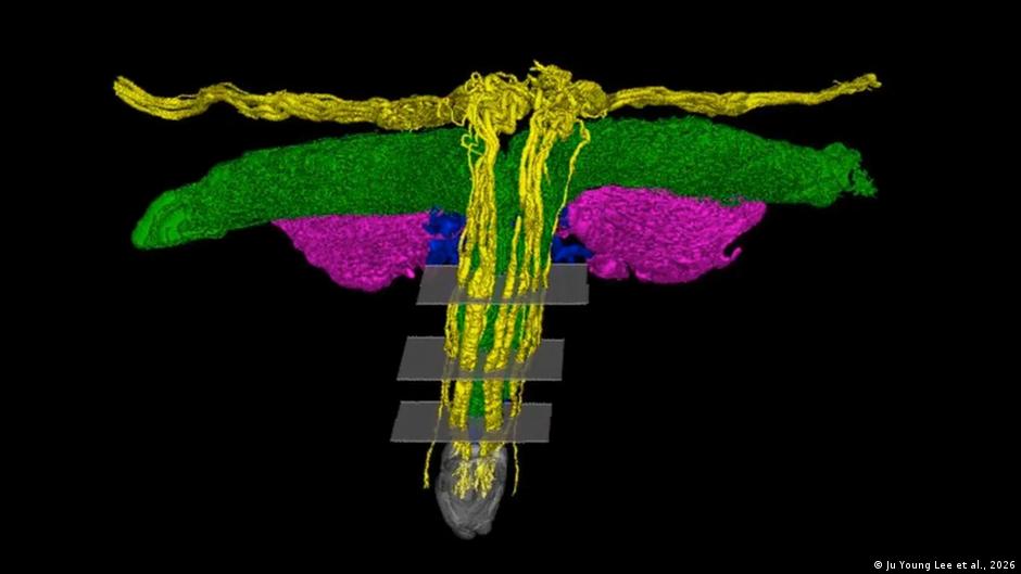
Mapping the clitoris in 3D
a new one 3D study from Netherlands Now it is helping to bridge some long-standing knowledge gaps. A research team led by neuroscientist Ju Young Lee at the Amsterdam University Medical Center examined two female bodies donated to science using synchrotron radiation – an exceptionally high-resolution form of X-ray imaging. The technology allows structures to be seen in microscopic detail. Traditional methods such as MRI can capture large anatomical features, but they are not able to completely reconstruct the fine neural pathways of the clitoris in three dimensions.
The images reveal how complex the clitoral nervous system really is. The researchers traced the three-dimensional course of the dorsal nerve of the clitoris, the organ’s primary sensory nerve, from the pelvis to the clitoris glands. Inside the glans penis, several large nerve trunks emerge in a tree-like branching pattern towards the surface – some measuring up to 0.7 millimeters in diameter.
Contrary to earlier beliefs, the nerves do not contract but form a complex, tree-like branching pattern. The images also show that some nerve branches extend beyond the glans to the clitoral hood and to the mons pubis, the fatty tissue over the pubic bone.
Independent experts say progress lies less in discovering a new structure than in finally seeing it in full detail. “For the first time, the full trajectory of the terminal nerve branches of the clitoris has been mapped in three dimensions,” Georga Longhurst, head of the department of anatomy and physiology at the University of London, told DW. “Previous dissection and MRI studies have revealed these nerves before, but never achieved this level of detail.”
An organ that was ignored for decades
One reason the clitoris has been neglected for so long is that, for decades, it was limited to its visible tip, when in reality, most of the organ is inside the body. This broader physiological understanding only began to enter the medical mainstream in the late 1990s and early 2000s.
A key figure in this change was the Australian urologist Helen O’Connell. Using MRI, she showed that the clitoris is not a small external “nub”, but a large and complex organ – measuring approximately 8 to 12 centimeters (3.1 to 4.7 in) in total length when including its internal structures. The visible glans is only the outer portion of a structure that extends beneath the pubic bone and surrounds the vaginal opening, composed of erectile tissue that fills with blood during arousal.
There is no focus of research on the clitoris yet
Joo Young Lee trained as a neuroscientist with a long-standing focus on the brain. However, in recent years, neuroscience has increasingly focused its attention on peripheral nervous systems, such as the gut. At a major European neuroscience conference, Lee once asked if anyone was studying how nerves in the gynecological organs communicate with the brain. One panelist’s response stuck with him: “Oh, I never thought about that.“
Lee couldn’t let the question go. After completing her PhD, she joined the Amsterdam University Medical Center – part of the International Human Organ Atlas HubA project that aims to systematically map the human body using synchrotron imaging. Think of it as a kind of Google Earth for human anatomy.
“Of course, the clitoris is one of the human organs,” Lee told DW, “so it was important to include it.”
Why does this matter to medicine?
Since the preprint was released, Lee says surgeons have already contacted him to say the findings are helpful in their daily work. She explains that knowing the detailed anatomy of the clitoris “will help surgeons operating on the vulvar area to avoid nerve damage.”
The study authors emphasize that the data may be particularly relevant to surgeries involving the vulva, such as childbirth, gender-affirming surgery and reconstructive surgery after genital mutilation.
How big the gap still is between research and clinical reality becomes clear when talking to Mandy Mangler, a senior gynecologist and obstetrician at a hospital in Berlin. When she saw the new images, she was impressed – not because they overturned everything she knew, but because they finally provided solid evidence.
“There has been very little scientific research on the clitoris,” Mangler told DW. “The idea that nerves extended to the mons pubis and labia was plausible – now this has finally been shown.”
This matters not only for surgery and sexual medicine, but also for the treatment of genital injuries. Mangler points out that the clitoris barely appears in medical education. As a result, doctors perform operations in the vulvar area without fully understanding the underlying nerve anatomy. Therefore pain, sensory loss or sexual dysfunction are often not linked to previous surgery or childbirth.
Penis, Clitoris and Gender Health Differences
Mangler makes a direct comparison to men’s health. In her hospital, she shares operating facilities with urologists.
“I see every day how much effort is put into protecting the nerves during penile surgery,” she says. “There’s research, training, and awareness. When it comes to the clitoris, no one cares.”
To Mangler, it’s a textbook example of the gender health gap — medical standards that are routine for men but missing for women, not out of malice but because of historical neglect. This is a topic he also addresses in his recent book “Don’t miss the clitoris.“
not the last picture
In public reports, the study has been described by some as the first “complete mapping” of the clitoral nerves. Lee clearly disagrees with that wording. Only two post-mortem samples from older women were examined. How the structure and function of the clitoris change across the lifespan – during puberty, pregnancy, menopause or the menstrual cycle – is largely unknown.
“As a scientist, it’s not possible to have the whole picture,” says Lee. “Future technologies will bring greater insight.” There are still many more puzzle pieces to be solved – and Lee hopes to find them. The new clitoral study has no endpoint.
Longhurst also says, “The field of clitoral science should expand.” “It shouldn’t be a special interest anymore.”
Mangler says there needs to be a change in the way doctors treat the clitoris.
“In every gynecological surgery and obstetric care, the anatomy and physiology of the clitoris must be considered and protected,” she says, “just like we do with the penis.”
Edited by: Carla Bleiker